Stadlmann 2006 Cell Biochem Biophys
| Stadlmann S, Renner K, Pollheimer J, Moser PL, Zeimet AG, Offner FA, Gnaiger E (2006) Preserved coupling of oxydative phosphorylation but decreased mitochondrial respiratory capacity in IL-1ß treated human peritoneal mesothelial cells. Cell Biochem Biophys 44:179-86. |
Stadlmann S, Renner K, Pollheimer J, Moser PL, Zeimet AG, Offner FA, Gnaiger E (2006) Cell Biochem Biophys
Abstract: The peritoneal mesothelium acts as a regulator of serosal responses to injury, infection, and neoplastic diseases. After inflammation of the serosal surfaces, proinflammatory cytokines induce an “activated” mesothelial cell phenotype, the mitochondrial aspect of which has not previously been studied. After incubation of cultured human peritoneal mesothelial cells with interleukin (IL)-1β for 48 h, respiratory activity of suspended cells was analyzed by high-resolution respirometry. Citrate synthase (CS) and lactate dehydrogenase (LDH) activities were determined by spectrophotometry. Treatment with IL-1β resulted in a significant decline of respiratory capacity (p < 0.05). Respiratory control ratios (i.e., uncoupled respiration at optimum carbonyl cyanide p-trifluoromethoxyphenylhydrazone concentration divided by oligomycin inhibited respiration measured in unpermeabilized cells) remained as high as 11, indicating well-coupled mitochondria and functional integrity of the inner mitochondrial membrane. Whereas respiratory capacities of the cells declined in proportion with decreased CS activity (p < 0.05), LDH activity increased (p < 0.05). Taken together, these results indicate that IL-1β exposure of peritoneal mesothelial cells does not lead to irreversible defects or inhibition of specific components of the respiratory chain, but is associated with a decrease of mitochondrial content of the cells that is correlated with an increase in LDH (and thus glycolytic) capacity. • Keywords: Peritoneal mesothelial cells, Interleukin-1β, Mitochondria, Respiration, Citrate synthase, Lactate dehydrogenase, Cell viability
• O2k-Network Lab: AT Innsbruck Gnaiger E
Labels: MiParea: Respiration, mt-Biogenesis;mt-density, Pharmacology;toxicology
Stress:Cell death Organism: Human Tissue;cell: Endothelial;epithelial;mesothelial cell Preparation: Intact cells
Regulation: Coupling efficiency;uncoupling, Substrate Coupling state: ROUTINE, ETS"ETS" is not in the list (LEAK, ROUTINE, OXPHOS, ET) of allowed values for the "Coupling states" property.
HRR: Oxygraph-2k
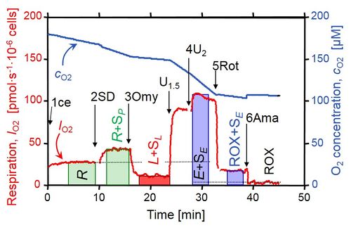
Coupling control protocol
- » CCP(S)01
| Step | <Symbol in 2006 publication> | Respiratory state | Pathway control | Comment |
|---|---|---|---|---|
| R | < E > | ROUTINE | ce | pce without substrate are in a state of ROX. |
| 1SD | < S > | R+SP | ROUTINE (R) + S (SP) | pce are in state SP. |
| 2Omy | < O > | L+SL | LEAK (L) + S (SL) | ce and pce are in the LEAK state. |
| 3U | < 3u > | E+SE | ETS (E) + S (SE) | ce and pce are in the ETS state. |
| 4Rot | < R > | ROX+SE | ROX+S (SE) | ce are in the ROX state, pce are in the ETS state. |
| 5Ama | < A > | ROXAma | ROX |
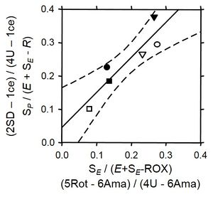
Cell membrane permeability index from the CCP
- These respiratory indices are based on the assumption that respiratory integrity of mitochondria is maintained after cell membrane permeabilization using the mitochondrial respiration medium MiR05.
- The labels of the axes show the respiratory states as steps (mark names), and respiratory states.
- The two indexes derived from (1) the succinate control step, and (2) the antimycin A control step are in close agreement (modified Fig. 2A from Stadlmann et al 2006).
Succinate control step
| Step | <Symbol in 2006 publication> | Respiratory state | Comment |
|---|---|---|---|
| 1SD-R | < S-E > | R+SP - R | Stimulation of respiration by succinate & ADP (SD) in permeable cells (pce) over endogenous ROUTINE respiration (R) in intact cells (ce). ce are not permeable to SD, and pce are depleted of substrates and adenylates. Hence SD stimulates mitochondrial respiration in pce only. |
| 3U – R | < 3u-E > | E+SE - R | Total capacity over endogenous ROUTINE respiration in permeable
and nonpermeable cells. All oligomycin-inhibited cells are stimulated by uncouplers (3U), because intact cell membranes are permeable for the uncoupler and respiration is supported by endogenous substrates in ce, whereas pce respire on succinate. |
- R+SP - R = SP (pce only)
- E-R is the excess E-R capacity, ExR (ce only)
- Normalization of the cell membrane permeability index
- SP / (ExR+SE)
- If all cells are viable (ce), then this index equals 0, since SP = 0.
- If all cells are permeable (pce), then this index equals 1, since SP and SE are practically identical in many cases, then SP/SE = 1.0.
Antimycin A control step
| Step | <Symbol in 2006 publication> | Respiratory state | Comment |
|---|---|---|---|
| 4Rot-5Ama | < R-A > | SE,mt (mt is ROX-corrected) | Succinate-supported respiration in pce (in the presence of the CI inhibitor
rotenone, Rot) over antimycin A–inhibited oxygen uptake (Ama) of all cells. This effect is specific for pce in the presence of succinate and uncoupler. |
| 3U-5Ama | < 3u-A > | (E+SE)mt (mt is ROX-corrected) | ETS capacity of ce plus SE of pce over antimycin A–inhibited respiration (mt; ROX-corrected). |
- SE - ROX = SE,mt (pce only)
- (E+SE) - ROX = (E+SE)mt
- Normalization of the cell membrane permeability index
- SE,mt / (E+SE)mt
- If all cells are viable (ce), then this index equals 0, since SE,mt = 0.
- If all cells are permeable (pce), then this index equals 1, since in this case Emt = E-ROX = 0.


